..:: klik untuk One 4 Share versi mobile ::.. ..:: Di sini, Rp 10.000,- bekerja untuk Anda ::..
Showing posts with label fisioterapi. Show all posts

Monday, September 5, 2011

4 Latihan Sederhana agar Hidup Lebih Lama

Berdasarkan penelitian Komunitas Kanker Amerika (American Cancer Society), para wanita yang duduk lebih dari enam jam sehari memiliki beresiko meninggal lebih dari 40% dibandingkan mereka yang duduk kurang dari 3 jam sehari.
"posisi kuda" Latihan Panjang Umur

Solusinya lakukan 4 Latihan Sederhana Berikut ini agar Hidup Lebih Lama / Panjang Umur:

Thursday, June 16, 2011

High Heels Mengancam Kaki Wanita

high heels ancam kaki wanita [img:telegraph]
Sebuah polling survey dari The Society of Chiropodists and Podiatrists menemukan bahwa sekitar seperempat wanita memakai high heels setiap hari, hal ini meningkatkan resiko mereka mengalami masalah serius pada kaki mereka.

High heels mempengaruhi postur tubuh wanita, saat mengenakan high heels beban pada pergelangan kaki dan lutut lebih berat. Ini menyebabkan tekanan pada jaringan kartilago dan memicu terjadinya osteoarhritis [peradangan pada sendi].

Saturday, June 11, 2011

Es dapat digunakan untuk Terapi | Terapi Es | Cryotherapy

Terapi Es [img: Corbis]
Terapi dengan menggunakan es atau disebut dengan Cryotherapy merupakan salah satu metode hidroterapi [terapi dengan memanfaatkan air ] yang menggunakan es sebagai medianya. Prinsipnya terapi es ini memanfaatkan suhu es yang rendah (dingin).

Thursday, May 26, 2011

Physiotherapy for Children [FREE eBook]

Physiotherapy for ChildrenEbook about Pediatric “Physiotherapy for Children”. Book Description taken from amazon, to download free ebook “Physiotherapy for Children”just click [FREE DOWNLOAD] at the end of post.

The Spastic Forms of Cerebral Palsy: A Guide to the Assessment of Adaptive Functions [FREE eBook}

The Spastic Forms of Cerebral Palsy: A Guide to the Assessment of Adaptive FunctionsEbook about Pediatric in Cerebral Palsy “The Spastic Forms of Cerebral Palsy: A Guide to the Assessment of Adaptive Functions”. Book Description taken from amazon, to download free ebook “The Spastic Forms of Cerebral Palsy: A Guide to the Assessment of Adaptive Functions”just click [FREE DOWNLOAD] at the end of post.

Early Diagnosis and Interventional Therapy in Cerebral Palsy [eBook FREE]

Early Diagnosis and Interventional Therapy in Cerebral Palsy: An Interdisciplinary Age-Focused Approach (Pediatric Habilitation)
Cerebral Palsy
Ebook about Pediatric in Cerebral Palsy "Early Diagnosis and Interventional Therapy in Cerebral Palsy". Book Description taken from amazon, to download free ebook "Early Diagnosis and Interventional Therapy in Cerebral Palsy" just click [FREE DOWNLOAD] at the end of post.

Monday, May 23, 2011

Stretch Exercises Increase Tolerance to Stretch in Patients With Chronic Musculoskeletal Pain: A Randomized Controlled Trial

Background: Stretch is commonly prescribed as part of physical rehabilitation in pain management programs, yet little is known about its effectiveness.

Objective: A randomized controlled trial was conducted to investigate the effects of a 3-week stretch program on muscle extensibility and stretch tolerance in patients with chronic musculoskeletal pain.

Proses Terjadinya Osteoporosis

Berikut ini video gratis tentang Osteoporosis / Tulang keropos / Pengeroposan tulang | Osteoporosis adalah | Proses terjadinya Osteoporosis | Osteoporosis Tulang | Proses Pengeroposan Tulang
Skeletal Fitness by Mirabai Holland Osteoporosis Prevention Bone Loading and Strength Training Exercises:A Workout For Bones and Bone Health for Boomers, Seniors, and Beginners
Proses Terjadinya Osteoporosis
Proses Osteoporosis : Osteoporosis terjadi ketika tubuh gagal membentuk tulang baru dalam jumlah yang cukup, atau ketika terlalu banyak tulang tua ( tulang yang lebih dahulu terbentuk ) diabsorbsi (diserap) kembali oleh tubuh, atau mungkin keduanya. Pada tulang normal yang sehat, remodeling tulang ( proses pembentukan tulang ) terjadi ketika osteoclast memakan jaringan tulang dan osteoblast datang membangun / mengisi lubang tulanng (yang telah dimakan osteoclast) dengan jaringan tulang yang baru.

Osteoporosis Process | How Osteoporosis Occurs ??

FREE Video about Osteoporosis | Osteoporosis Process | How Osteoporosis Occurs ?
Your Bones: How You Can Prevent Osteoporosis & Have Strong Bones for Life Naturally
Osteoporosis Process
Osteoporosis occurs when the body fails to form enough new bone, or when too much old bone is reabsorbed by the body, or both. In normal healthy bone, remodeling occurs when osteoclasts eat away at the bone tissue and osteoblasts come in and refill the holes with new bone.

Saturday, May 21, 2011

LATIHAN SEDERHANA ATASI NYERI PUNGGUNG BAWAH [VIDEO GRATIS]

Postur tubuh yang kurang bagus karena otot-otot mengalami kelemahan seringkali menjadi penyebab atau memperburuk nyeri punggung bawah, tapi latihan-latihan yang dapat mengulur / merenggangkan otot-otot adduktor dapat meningkatkan komunikasi saraf dengan otot-otot pantat dan dapat membantu memperkuat punggung. Video GRATIS latihan ini dapat membantu mengurangi NYERI PUNGGUNG.

[ FREE VIDEO] EXERCISE FOR LOW BACK PAIN

Poor posture from slack muscles often causes or worsens LOWER BACK PAIN, but exercises that stretch the adductors, improve nerve communication to the gluteal muscles and reinforce the spinal arch can help. Reduce BACK PAIN with this FREE VIDEO of exercises to train PAIN - FREE.

Friday, May 20, 2011

Motor Control Exercise for Chronic Low Back Pain: A Randomized Placebo-Controlled Trial

[Background] The evidence that exercise intervention is effective for treatment of chronic low back pain comes from trials that are not placebo-controlled.

[Objective] The purpose of this study was to investigate the efficacy of motor control exercise for people with chronic low back pain.

Thursday, May 19, 2011

Effects of Mastectomy on Shoulder and Spinal Kinematics During Bilateral Upper-Limb Movement.

[Background ] Shoulder movement impairment is a commonly reported consequence of surgery for breast cancer.

[Objective] The aim of this study was to determine whether shoulder girdle kinematics, including those of the scapula, spine, and upper limb, in women who have undergone a unilateral mastectomy for breast cancer are different from those demonstrated by an age-matched control group.

Wednesday, May 18, 2011

Muscle Activation and Perceived Loading During Rehabilitation Exercises: Comparison of Dumbbells and Elastic Resistance


[Background]
High-intensity resistance training plays an essential role in the prevention and rehabilitation of musculoskeletal injuries and disorders. Although resistance exercises with heavy weights yield high levels of muscle activation, the efficacy of more user-friendly forms of exercise needs to be examined.

[Objective]
The aim of this study was to investigate muscle activation and perceived loading during upper-extremity resistance exercises with dumbbells compared with elastic tubing.

Tuesday, May 17, 2011

Progressive Resistance Training Improves Overall Physical Activity Levels in Patients With Early Osteoarthritis of the Knee: A Randomized Controlled Trial

Prescription of resistance training (RT) exercises is an essential aspect of management for knee osteoarthritis (OA). However, whether patients with knee OA who are randomly assigned to receive RT simply substitute RT for other modes of physical activity remains unclear.

The aim of this study was to determine the effect of a structured RT intervention on overall levels of moderate- and vigorous-intensity physical activity (MVPA) in patients with early-onset knee OA. The study compared patients with early-onset OA who participated in an RT program, those who participated in a self-management (SM) program, and those who participated in both RT and SM. Because participants randomly assigned to receive the RT intervention may simply switch activity modes, resulting in little net effect, we assessed total MVPA in addition to tracking changes in strength (force-generating capacity).

Wednesday, June 16, 2010

Waspadai Nyeri Pantat [ Sindroma Piriformis | Piriformis Syndrome ]


Sampai saat ini, belum ada yang tahu pasti apa yang menyebabkan sindroma piriformis. Beberapa ahli percaya bahwa sindroma piriformis / piriformis sindrom adalah sebutan untuk nyeri pada pantat / paha yang belum didiagnosis secara tepat ( jadi semacam keranjang sampah seperti halnya frozen shoulder ). Akan tetapi ada pendapat lain yang menyatakan bahwa sindroma piriformis merupakan gangguan yang nyata yang menyebabkan nyeri dan disability (Cluett, 2004).

Piriformis sebenarnya adalah nama otot yang terletak di belakang / sebelah posterior sendi panggul (hip joint). Otot piriformis sendiri merupakan sebuah otot kecil bila dibandingkan dengan otot-otot tungkai yang lain. Fungsi otot piriformis ini adalah sebagai penggerak aktif gerakan external rotasi sendi panggul ( hip joint) (Cluett, 2004). Selain itu otot piriformis juga berfungsi dalam menjaga keseimbangan ketika salah satu kaki terangkat dan sebagai stabilisator aktif daerah pelvic (Maggs, 2010).

Otot piriformis di inervasi oleh n. sciatic / n. ischiadicus (salah satu nervus terbesar tubuh manusia yang menginervasi ekstremitas bawah). Tendon otot Piriformis dan n. ischiadicus saling bersilangan di posterior sendi panggul, di bagian dalam pantat ( cluett, 2004). Atau dengan kata lain otot piriformis ini berada di bawah (di sebelah anterior) otot gluteus maximus.


Pada sindroma piriformis, terjadi semacam penjepitan n. ischiadicus oleh otot piriformis, sehingga menyebabkan n. ischiadicus teriritasi. Hal tersebut terjadi apabila otot piriformis memendek, sehingga n.ischiadicus terjebak. Akibatnya aliran / suplai darah ke . ischiadicus pun terhambat, sedangkan iritasi terjadi akibat tekanan oleh otot piriformis tersebut ( Cluett, 2004).

Maggs (2010) berpendapat bahwa salah satu penyebab sindroma piriformis adalah cedera. Otot piriformis sangat rentan untuk terjadi cedera berulang akibat gerakan (repetitive motion injury / RMI). RMI terjadi apabila otot bekerja diluar kemampuannya, atau tidak diberi cukup waktu untuk fase recovery, akibatnya, otot menjadi memendek (Maggs, 2010)

Gejala sindroma piriformis antara lain:
Pertama, nyeri di dalam dan di sekitar tulang panggul (Hip). Pemendekan otot meningkatkan tekanan diantara tendon dan tulang, sehinggga secara langsung terjadi ketidaknyamanan dan nyeri. Atau dapat juga hal tersebut menyebabkan bursitis (Maggs, 2010)
Kedua, nyeri pada bagian tengah pantat / bokong. Nyeri ini dapat di picu dengan memberikan tekanan pada daerah pantat (Maggs, 2010).
Ketiga, sindroma piriformis mungkin terjadi akibat ischialgia (Maggs, 2010).


Sindroma Piriformis juga sering disebut “deep buttock pain”. Hal itu karena nyeri yang di arasakan penderita berada jauh di dalam pantat. Penyebab lain yang dapat mengakibatkan nyeri tersebut antara lain karena masalah yang terjadi pada spinal ( tulang belakang), termasuk di dalamnya HNP, spinal stenosis, dsb. Selain itu juga dapat terjadi akibat tendonitis (Cluett, 2004)

Diagnosis sindroma piriformis diberikan apabila semua diagnosis tersebut tidak sesuai sebagai penyebab nyeri. Tanda-tanda lain dari sindroma piriformis dapat diketahui dengan pemeriksaan khusus untuk mengisolasi fungsi otot piriformis, dan mencari hal-hal yang menyebabkan nyeri pada otot piriformis (Cluett, 2004).


Sedangkan diagnosis Fisioterapi, impairment pada sindroma piriformis antara lain nyeri, spasme otot piriformis, pemendekan otot piriformis. Tiga hal tersebut, Nyeri , spasme dan pemendekan otot piriformis saling terkait satu sama lain sebagaimana telah dijelaskan di atas. Tentang nyeri sendiri dapat di baca di NYERI (PAIN). Selain tiga hal tersebut, apabila n. ischiadicus terjadi cedera, maka akan dapat menimpulkan gangguan baik motorik mapun sensorik area-area yang diinervasi oleh n. ischiadicus. Hal tersebut tentunya tergantung bagian mana yang mengalami cedera.

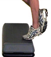
Limitasi fungsi akibat sindroma piriformis sendiri terjadi sebagai akibat langsung dari pemendekan otot piriformis, diantaranya kurangnya lingkup gerak sendi external rotasi hip. Di samping itu juga bias merupakan akibat tak langsung dari nyeri, dimana secara psikologis penderita nyeri akan membatasi / mengurangi aktivitas atau geraknya, sehingga berakibat pada fungsi ekstremitas bawah.

Penanganan Fisioterapi pada Sindroma Piriformis dapat menggunakan berbagai modalitas Fisioterapi untuk meringankan keluhan nyeri, dan sejauh mungkin mengatasi penyebab sindroma piriformis sendiri. Mengatasi penyebab sindroma piriformis, dalam hal ini yang dimaksud adalah penyebab berupa spasme dan pemendekan otot piriformis seperti dijelaskan di atas. Modalitas Fisioterapi yang dapat digunakan antara lain adalah Short wave diathermy (SWD), TENS, Ultra sound (US), massage (transfers friction), terapi latihan ( stretching otot piriformis), dsb.


Referensi:
Cluett, J. 2004. Piriformis Syndrome. http://www.about.com
Maggs, T.J. 2010. Piriformis Syndrome. http://www.spineuniverse.com


Disusun oleh : Rohmat Saputro Wibowo
Untuk mengutip artikel ini:
Wibowo, R.S., 2010. Sindroma Piriformis / Piriformis Syndrome. http://one4share.blogspot.com/


PEMERIKSAAN FISIOTERAPI PADA RUPTUR TENDI ACHILLES

pemeriksaan Fisioterapi RUPTUR TENDO ACHILLES
Anamnesis:
• Keluhan
- Nyeri di daerah pergelangan kaki, kadang hingga ke betis dan kaki
- Tidak dapat atau kurang mampu menggerakan kaki (terutama gerakan plantar fleksi)
- Kaku di pagi hari


Inspeksi
• Pembengkakan di daerah pergelangan kaki
• Deformitas / perubahan bentuk

Palpasi

Palpasi Ankle• Lokasi Tenderness / nyeri tekan, di tendo achilles
• Temperatur lokal
• Spasme otot? Terutama m. gastrocnemius


***

***


Pemeriksaan khusus

• Thompson’s test
thompson's test


• Squeeze test
Squeeze test


Pemeriksaan fungsional

Pemeriksaan fungsional dasar
• Calf raises (jinjit dengan kedua kaki)
• Single calf raise (jinjit dengan satu kaki)


Lain-lain (berkaitan dengan evaluasi)
• Nilai nyeri
Perlu diukur 3 jenis nilai nyeri, nyeri diam, nyeri gerak dan nyeri tekan. Penilaian dapat menggunkan VDS, VAS, maupun NRS.

• Lingkup Gerak Sendi
Dapat menggunakan goneometer, baik pasif maupun aktif. Nilai LGS normal dorsi-plantar flexi : S 20-0-45

• Kekuatan otot (MMT)
Perlu mengukur nilai kekuatan group otot dorsal flexor, plantar flexor, inversor, dan eversor.


Sunday, April 11, 2010

Acute Pain Management (3)

Pain Assessment

Pain History
The elements of a pain history provide information that can alert to the presence of a serious underlying condition. It is important to note that in the absence of a serious cause for the pain (e.g. fracture), it is not necessary to obtain a specific patho-anatomic diagnosis to manage acute musculoskeletal pain effectively.

Site
The anatomical site where the person feels the pain may or may not be the site of origin as in the case of referred pain. The clinician should ask which part hurts the most and whether the pain started there or elsewhere.

Distribution
The regions in which pain is felt should be described. Even a person who initially complains of ‘pain all over’ can usually describe distinct region(s) of pain (possibly large and overlapping). Having the patient draw their pain focus and radiation on a pain diagram clarifies its distribution and can act as a baseline from which to assess response to treatment and changes in pain patterns.

***


***

Quality
The quality of pain may be described in different ways. Somatic pain is usually deep, dull and aching. Radicular pain is mostly sharp and ‘electric’ or ‘shooting’. Neuropathic pain is often ‘burning’. Visceral pain is dull at first but sharp when lining tissues such as the peritoneum become involved.

Duration
By convention, pain present for less than three months is described as ‘acute’ pain. Chronic pain refers to pain present for greater than three months duration. Pain duration will affect pain management.

Temporal Factors
Pain may be constant or intermittent. If pain is constant the history should elicit whether its intensity varies. If pain is intermittent, the history should elicit its pattern in relation to time
of day, activity and duration.

Intensity
The intensity of pain reflects the impact of the experience, not necessarily the degree of nociception. Even though pain is essentially subjective (Merskey and Bogduk 1994) it is important to assess the intensity of the pain. Simple tools can be used to assess pain at the initial and follow-up visits to evaluate progress. There is good correlation between the various types of
scales (Jensen at al. 1986). The Numerical Rating Scale is suitable for many clinical situations because it is simple to apply.

Aggravating and Relieving Factors
Aggravating factors include those that precipitate or worsenpain. Relieving factors are those that alleviate, reduce or abolish pain. People who say that nothing eases the pain can be asked about the posture in which they are least uncomfortable.

Impact on Activities of Daily Living and Sleep
The effects of pain on activities of daily living (ADL) determine
associated disabilities and handicaps (WHO 1986)
Identifying such effects gives the clinician an idea of the impact of pain on the patient’s lifestyle. The effect of pain on sleep should be specifically sought; sleep deprivation is a powerful amplifier of the pain experience.

***


***
Associated Symptoms
These include any symptom apparently associated with the painful condition, in contrast to symptoms associated with other conditions the person may also have.

Onset (Precipitating Event)
The first appearance of the pain and the circumstances in which it started should be assessed. The clinician should distinguish between an event that may have aggravated rather than
precipitated the pain.

Previous Similar Symptoms
Previous experience of similar symptoms suggests a recurrent condition.

Previous Action to Relieve Pain
All measures used for the condition before (and their effectiveness) should be noted. Unwanted effects associated with past treatment should also be recorded. Information on how each
intervention was applied can be helpful, as treatment ‘failures’ may be due to misapplication rather than to true failure of effect.

Current Action to Relieve Pain
All forms of treatment in current use should be noted. The clinician should ask about the use of physical interventions, including self-applied measures, all passive treatments, and all substances whether prescribed or otherwise that the person is taking or applying, with an appraisal of the helpfulness of each.

Acute Pain Management (2)

Acute Pain

The term ‘acute pain’ refers to pain that has been present for less than three months (Bonica 1953; Merskey 1979). Successful management of pain in the acute phase is essential to prevent transition to chronic pain, which presents a significant individual, social and financial burden. Chronic pain is pain that has been present for longer than three months (Merskey and Bogduk 1994).

The NHMRC (1999) cites a number of misconceptions about the management of acute pain, including a lack of understanding of the pharmacokinetics of analgesics, mistaken beliefs about addiction, poor knowledge of dosage requirements, concerns about side effects and the concept that pain is not harmful.

***


***

Factors Influencing the Progression from Acute to Chronic Pain

Individuals vary in their potential to develop chronic pain. A combination of behaviours, beliefs and emotions may be involved in the transition from acute to chronic pain (Linton 2002). When pain is unrelieved over time, or if there are recurrent episodes of pain, persistent pain may develop.

The development of chronic pain is likely to be the result of small, cumulative changes in lifestyle that have been made to cope with acute musculoskeletal pain (Linton 2002). The intensity, duration and character of the pain influence the psychosocial response and the psychosocial response in turn influences the course of events.

There is strong evidence that psychosocial factors at work (i.e. occupational factors) are tied to the development of chronic pain. Job satisfaction may protect against the progression from acute low back pain to chronic low back pain. It is essential to identify those at risk of developing chronic pain and to intervene early to prevent this occurrence.


taken from:
Australian Acute Musculoskeletal Pain Guidelines Group;2003; Evidence-based Management of Acute Musculoskeletal Pain; Bowen Hills: Australian Academic Press Pty. Ltd.


Wednesday, February 24, 2010

Waktu optimal TENS untuk pengurangan nyeri pada OA lutut

Osteoarthritis (OA) adalah salah satu bentuk arthritis yang paling sering terjadi (memiliki angka prevalensi yang tertinggi dibandingkan jenis arthritis lain). Sebagian besar penderita OA adalah para lansia dengan usia 65 tahun keatas terutama OA lutut.

Terapi yang efektif untuk menangani nyeri (yang biasanya merupakan keluhan utama penderita OA) akan dapat meningkatkan kualitas mobilitas penderita. Salah satu modalitas Fisioterapi untuk mengontrol nyeri yang sering diaplikasikan adalah TENS. TENS dalam berbagai penelitian telah terbukti efektif menurunkan nyeri pada penderiata OA lutut, tapi beberapa hasil penelitian masih kontroversial.


***


***


Dalam berbagai penelitian yang telah dilakukan, ternyata masih belum diketahui berapa waktu terapi yang optimal pengaplikasian TENS pada penderita OA lutut. Dalam peneltian sebelumnya waktu yang digunakan bervariasi antara 30 hingga 60 menit per hari.

Cheing et al (2003) dalam penelitiannya membahas hal tersebut (waktu yang paling efektif untuk pengaplikasian TENS pada penderita OA lutut). Cehing et al (2003) membandingkan empat model pengaplikasian TENS yaitu TENS dengan lama pengaplikasian 20 menit, 40 menit, 60 menit dan TENS plasebo. TENS yang digunakan berupa TENS konvensional dengan frekuensi 100 pps, durasi pulsa 200 mikrosecon, dengan penempatan electrode pada titik-titik akupuntur di daerah lutut.

Hasil penelitian Cheing et al (2003) tersebut adalah bahwa TENS konvensional pada penderita OA lutut paling efektif (waktu optimal) diaplikasikan selama 40 menit.


Baca (download):

Cheing, G. L. Y. et al, 2003, Optimal Stimulation Duration of TENS In The Management of Ostheoarthritic Knee Pain, J Rehabil Med 35: 62–68


..::klik untuk One 4 Share versi mobile ::.. _ __